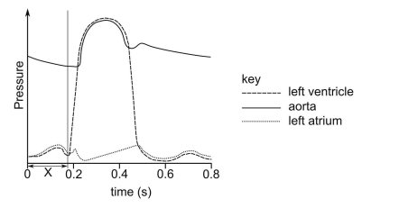
The diagram shows how the pressure changes in the left atrium, left ventricle and the aorta during one heartbeat in a healthy human.
Which row shows the correct events during time period X?
A. row 2
B. row 3
C. row 5
D. row 4
E. row 1
The diagram shows how the pressure changes in the left atrium, left ventricle and the aorta during one heartbeat in a healthy human.
Which row shows the correct events during time period X?
A. row 2
B. row 3
C. row 5
D. row 4
E. row 1
To understand the diagram in the question, we need to understand the pressure changes during a complete cycle of one heartbeat in different parts of the heart.
Instead of analyzing the entire diagram, let’s try to break down each section in turn.
The left ventricle is the chamber that pumps oxygenated blood to the entire body. Transferring blood to the whole body requires pumping against the resistance of many blood vessels. This is why the cardiac muscle of the left ventricle is wider than the muscle of the right ventricle, which pumps blood from the heart to the lungs.
By definition, pressure = force/area . We should try to apply this concept to our question to make it easier to remember.
The force in our case is the blood that “clashes” against the left ventricle during the contraction.
Taking the same amount of particles (in our case, blood) while decreasing the volume of the container these particles are found in will lead to an increase in pressure as there are more particles clashing with force against the container (area).
We can expect an increase in pressure of the ventricles during rapid filling and contracting. Rapid filling of the right and left ventricles co-occurs (as per the IMAT), and when the ventricles contract, the atria will always relax. The atria and the ventricles won’t contract at the same time.
To allow the aortic valve to open, the heart must contract until it reaches a point where the pressure of the ventricle is more significant than the pressure in the aorta; otherwise, the valve won’t open.
Once the aortic valve opens, the pressure of the ventricle and the aorta equalizes until the pressure of the ventricle becomes less than the pressure of the aorta.
Then, as seen in the diagram, we see a slight backflow in the aorta back to the heart, which will cause blood to flow to the cardiac muscle through the cardiac arteries.
The process we describe seems to occur after 0.2s on our diagram, the pressure of the left ventricle increases until it equalizes with the aorta. Before this process, we must fill the ventricles with blood. The atrium contracts (with the help of gravity) and allows blood to go to the ventricles while the ventricular muscles relax.
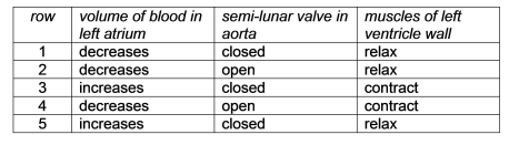
The blood volume in the left atrium will decrease while the blood volume in the ventricle increases, and the semilunar valve will be closed. In contrast, the mitral valve will stay open to allow the blood to flow from the left atrium to the ventricle, and the muscles of the left ventricle must relax to allow the ventricle to get filled.
The answer must be E, row 1.