Can you please explain about the structure of the centrioles? i understand its 9 triplets of microtubules but i don’t understand the shape its forming, i can’t visualize it.
Great question! Understanding the structure of centrioles can be a bit challenging, but I hope the explanation and images below will help you visualize it better. And don’t worry, we will be discussing this in more detail during the next 2-3 classes as the 3rd slide is about this exact thing.
Centrioles are tiny cylindrical organelles composed mainly of a protein called tubulin. They are found in most eukaryotic cells and play an essential role in cell division and the formation of cilia during interphase. You can read more about this here.
The typical structure of a centriole is composed of nine sets of short microtubule triplets, arranged in a cylinder. Each triplet is made up of three concentric rings of microtubules that form together. This structure makes them very strong. Each microtubule in a triplet is made up of small units of tubulin, a small monomer that can join together to create long, hollow tubes that resemble straws. You can find more details here.
Here’s an image that should help you visualize the structure of a centriole:
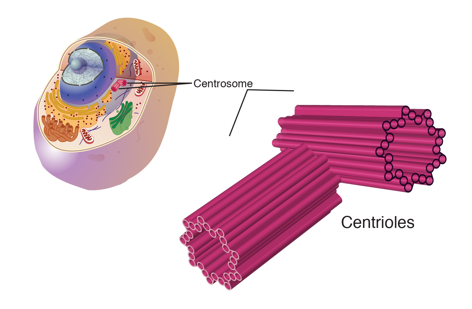
As you can see from the image, the nine triplets of microtubules form a cylinder. Think of it as a bundle of straws tied together, where each straw is a triplet of microtubules. Each triplet is bonded together by special proteins that give a centriole its shape.
I hope this helps! We’ll be diving deeper into this topic in our upcoming classes.